Imaging Tests for Lobular Breast Cancer
Lobular breast cancer is cancer that starts in the lobules, the glands in the breast that produce milk. Lobular breast cancer is the second most common breast cancer, accounting for about 10% of breast cases in the United States. Lobular breast cancer doesn’t always form a lump and can spread to areas of the body different from ductal breast cancer, such as the abdominal lining or the tissue around the kidneys or the eyes.
Listen to the podcast to hear Dr. Jochelson explain:
- Sponsor Message
why lobular breast cancer is harder to see on mammograms
- Sponsor Message
the imaging tests used to figure out if lobular breast cancer has spread to other areas of the body
- Sponsor Message
the difference between a FES PET scan and a FDG PET scan
Welcome to The Breastcancer.org Podcast, the podcast that brings you the latest information on breast cancer research, treatments, side effects, and survivorship issues through expert interviews, as well as personal stories from people affected by breast cancer. Here’s your host, Breastcancer.org senior editor Jamie DePolo.
Jamie DePolo: Hello. Thanks for listening. Our guest today is Dr. Maxine Jochelson, chief of the Breast Imaging Service at Memorial Sloan Kettering Cancer Center. Besides specializing in breast imaging, she also has expertise in interpreting PET/CT scans. Dr. Jochelson joins us to discuss imaging tests for invasive lobular breast cancer, including telling us why lobular breast cancer is more difficult to see on mammograms. Dr. Jochelson, welcome to the podcast.
Dr. Maxine Jochelson: Thank you for having me.
Jamie DePolo: I know we're going to learn a lot today, and just in case anybody listening isn't aware, lobular breast cancer is cancer that starts in the lobules, the glands in the breast that produce milk, and lobular breast cancer is the second most common breast cancer, accounting for about 10% of breast cancer cases in the United States. Ductal breast cancer, which starts in the ducts, which are the tubes that carry milk from the lobules to the nipple, is more common, and that makes up about 80% of cases.
So, Dr. Jochelson, I've read that, in many cases, lobular breast cancer doesn't cause a lump. Is this why it's harder to see on a mammogram?
Dr. Maxine Jochelson: This is why it's harder to see on a mammogram and why, by the time the patient does have a symptom, it's often later because, you know, sometimes the patient will find a small lump with other cancers, and in this case, it doesn't always happen.
Jamie DePolo: Okay, and when you're reading a mammogram and you see a suspicious area, do you automatically recommend another type of imaging, or does it depend on what you see? How does that work?
Dr. Maxine Jochelson: So, first of all, there are two kinds of mammograms. There are screening mammograms, and those are mammograms that are done in women who have no symptoms at all, and then there are diagnostic mammograms, where a woman may come in with a symptom. And we approach them slightly differently.
The patient who has a symptom, if she has a lump, the technologist has marked it off and said, you know, Mrs. Jones has a lump here in the 5 o'clock axis. I look at the mammogram. We do tomosynthesis on almost all of our patients, which is kind of like a CT scan through the breast, and so, that actually prevents us from doing an additional spot film, which we used to do. So, we take a look at the mammogram. If I don't see anything on the mammogram that goes with that lump, I will then do an ultrasound.
[The] other group of patients are screening patients, and they're asymptomatic, and we read those...so, the diagnostic, we read while the patient is in the building. The screening, we batch read, and we read it in lumps so that the patient gets the results by the next business day, and then, if I see something abnormal...and the same with the diagnostics. If I see something else abnormal, then I will do additional mammographic imaging.
Jamie DePolo: Okay. Okay, and just so everybody knows, too, I think, in a lot of places, tomosynthesis, that you mentioned, is called a 3D-mammogram, so it takes a 3D image of the breast.
Dr. Maxine Jochelson: So, it actually isn't a 3D image, but we call it a 3D. That's the irony of it. That's why I call it tomo...because it's really not three-dimensional, but that's the best we could come up with so people don't have to say tomosynthesis, which is hard enough.
Jamie DePolo: Okay. So, it's aiming to be a 3D mammogram?
Dr. Maxine Jochelson: Exactly.
Jamie DePolo: Okay. Okay. Okay. So, if you're...say you're looking at a diagnostic mammogram and lobular breast cancer might be suspected, and I know if there's no lump, like, what do you do then? How do you approach that?
Dr. Maxine Jochelson: You know, when I read any mammogram, I'm just looking for a cancer. You know, in the back of my brain, although, yes, there are small groups of women who have a likelihood of getting ILC because of a genetic thing, that's not that common. So, as...whenever I open up a mammogram in an asymptomatic woman, or symptomatic, I'm looking at it for any kind of cancer.
I don't really think, “Oh, I'm looking for invasive lobular,” but there are patterns that you do see when you have...and most of the time, that this is a more advanced lobular cancer, and that is you see an asymmetry. The breast looks a little different than the breast...than the other breast, for example. Or there's a well-known syndrome when it's fairly advanced, is that you have a shrinking breast. So, if you see old films and you see, all of a sudden, one breast is getting smaller, those are the kinds of things.
But by the time you're seeing those things, the tumor's been there for a while, but those are the things that makes your brain go lobular versus ductal cancer, but other than that, if it's a symptomatic patient and they don't feel a lump, but there's a ridge of tissue, sometimes that's the description. It's just some thickening or things like that, but that's very nonspecific, and you can have that from an invasive ductal cancer or from nothing and things like that. So, I still approach every case just looking for a breast cancer.
Jamie DePolo: That makes sense. So, if there is no lump...I'm trying to understand in my brain, like, how...I guess I'm wondering, if lobular breast cancer isn't very advanced, is it possible to catch earlier, or because it really doesn't often show symptoms, are they mostly caught later? I'm trying to understand that process.
Dr. Maxine Jochelson: Well, you can find a lobular cancer early. Sometimes, because it does disrupt the architecture of the breast, you know, and it may just be a small area early on, and one of the things that tomo is very good for is looking at what we call asymmetries or architectural distortion, because since you're looking at such thin slices through the breast.
You know, in the old days, the whole breast was sitting there, you know, in a 2D image, and you see everything on top of everything. Whereas, with this, you are going in thin sections through the breast, and so, a subtle area of change, you can detect more easily than you could on the old type of mammograms. So, you can find them early. Sometimes they present with microcalcifications, in which case, those are things that we find either way, as well.
Jamie DePolo: Okay, and I wanted to ask you, too, I know sometimes PET scans or CT scans are recommend to sort of help with the diagnosis of lobular invasive breast cancer. Some members of our community who've been diagnosed with lobular breast cancer have talked about, you know, what's the difference between an FES-PET scan versus an FDG-PET scan? And how some were recommended to get one versus the other. So, if you could kind of explain those for us?
Dr. Maxine Jochelson: I just want to make it clear, this is not to make the diagnosis in the breast.
Jamie DePolo: Oh, okay.
Dr. Maxine Jochelson: PET scanning is not a tool that we use to look for cancer in the breast. What we're talking about is a woman who has been diagnosed with invasive lobular breast cancer, okay, and then we're looking for metastatic disease.
Jamie DePolo: Oh, I see. Okay.
Dr. Maxine Jochelson: Okay? And so, that's what we call systemic staging. So, we systematically stage women when they have a more advanced breast cancer at presentation, if they have no symptoms, okay? So, classically, you know, we stage women I through IV. One is just a small breast cancer, no adenopathy. Two, the breast cancer's a little bit larger and may have abnormal lymph nodes. Three is more locally advanced, and IV is metastatic, okay?
So, stage I patients, we do not systemically stage. There's IIA and IIB. IIA we don't systematically stage, but we are moving towards systemically staging women with a bigger cancer and axillary nodes. The tools that we used have changed over a period of time. We used to just use CAT scans and bone scans, but we did find out, once we started doing PET, that PET was more sensitive and more accurate. So, PET, it's FDG-PET, and FDG is a sugar. You know, it's a sugar tab, and since cancers need sugars to grow...I mean, this is very simplistic.
So, we add a tracer to the sugar, and then the patient lies still for an hour after they've done this, and they have to fast beforehand, and then, while they're resting, the FDG will go to areas of cancer. It can also go to areas that are not cancer. For example, the heart, because the heart's beating, but that is the reason we're doing it, and it will show cancers not so much in the brain, but every place else in the body. What we've learned, over a period of time, FDG is fantastic for invasive ductal cancers.
The more aggressive cancers take it up even faster, and it's also used in every other...in not just breast cancer. The more aggressive cancers will be...the uptake is hot, it gets very high. You see a lot of uptake, and that's a really great way to stage and follow women with breast cancer. After a while, we began to realize that women with invasive lobular cancers don't necessarily take up the FDG at all or to the same extent as women with invasive ductal cancers, and over a period of time, we have looked at other tracers, okay?
So, one of them is called FACBC. It was actually a tracer that was also good in prostate cancer. It was good, but it was not perfect. But more recently, FES, so, it's an estradiol, it’s kind of a subset of estrogen, and this is an...the E in FES stands for the estradiol, and so, it's a test that's similar to a PET scan, in the sense that you get an injection of this tracer, but you don't need to lie still or whatever, and then you'll be scanned. And what we've learned is that, in breast cancers that are less aggressive, so that includes...or slower growing, So, that includes invasive lobulars. But some estrogen receptor-positive cancers that are not lobular cancers, but are not as aggressive as others, then FES is better in those specific cancers than FDG.
Now, this is a relatively newly FDA-approved tracer. It has one flaw, and that is it kind of...it goes to the liver, and so, the liver is so hot, that you can't necessarily find metastases in the liver like you can on an FDG. FDG doesn't...you can't see the brain very well. With FES, you can.
Otherwise, it shows you the rest of the body beautifully, and in fact, we have certainly seen patients in whom the FDG is negative or not so...you know, not so much going on, and the FES is very...it's FES-avid, so you see the tumor well. At this point in time, because this is still relatively new, in most women, we start with the FDG PET/CT, because that still is the standard of care, and in many women with ILC, it's great. So, it's not useless to do that, but if we are still suspicious, then we do the FES scan.
Jamie DePolo: Okay. So, somebody could have both?
Dr. Maxine Jochelson: They may have to adjust until we have more experience with FES.
The other thing that's very interesting, it's not only that it shows tumor. By showing the FDG scan and the FES scan, it can also tell you which drugs are more likely to work in your tumor. So, if the FES does not take up a lot...if you don't see a lot of uptake, then those women are less likely to get a benefit from certain hormone therapies than women where there's more uptake, because the FES is telling you about the hormonal milieu in your cancer. So, it helps you predict response to treatment by certain drugs and also comparing to FDG.
If the patient has more FDG uptake, it's a more aggressive tumor, and so, those patients don't necessarily do as well as a patient with FES. So, there's all these...and these are little things we're learning. So, it's not just finding the cancer. It's telling you how it should be treated and how it will behave.
There is one more tracer that's on the market soon called FAPI, which may actually be better than anything, but we don't have it FDA approved yet, and we're always looking for better and better ways of finding these cancers.
Jamie DePolo: Okay, thank you. That was very interesting. This new one that's not approved yet, has it been studied in breast cancer? I'm assuming it has?
Dr. Maxine Jochelson: Yes.
Jamie DePolo: Yes? Okay.
Dr. Maxine Jochelson: Yes, but it's not...you know, we certainly don't have the numbers of studies yet to do it, but I think it's going to be very good.
Jamie DePolo: So, if somebody that had been diagnosed with lobular breast cancer and it was suspected that it had metastasized, you know, maybe there are some other symptoms. From what you said, I just want to make sure I understand. It's likely the person would probably get the FDG-PET scan first, and then, if that didn't show anything, they would get the FES-PET scan? Is that how it works usually?
Dr. Maxine Jochelson: At this point in time. Again, I do believe that, within a short period of time, maybe a year or so, then they may get rid of the FDG, but right now, we need to use the tool that we know is really good and then grow from it. So, yes, for the time being, most places will do that.
Jamie DePolo: Okay. Okay, and then, I guess I'm wondering, too, is PET scan the primary type of imaging somebody would have, if they did have metastatic lobular breast cancer, to sort of monitor how treatment was working?
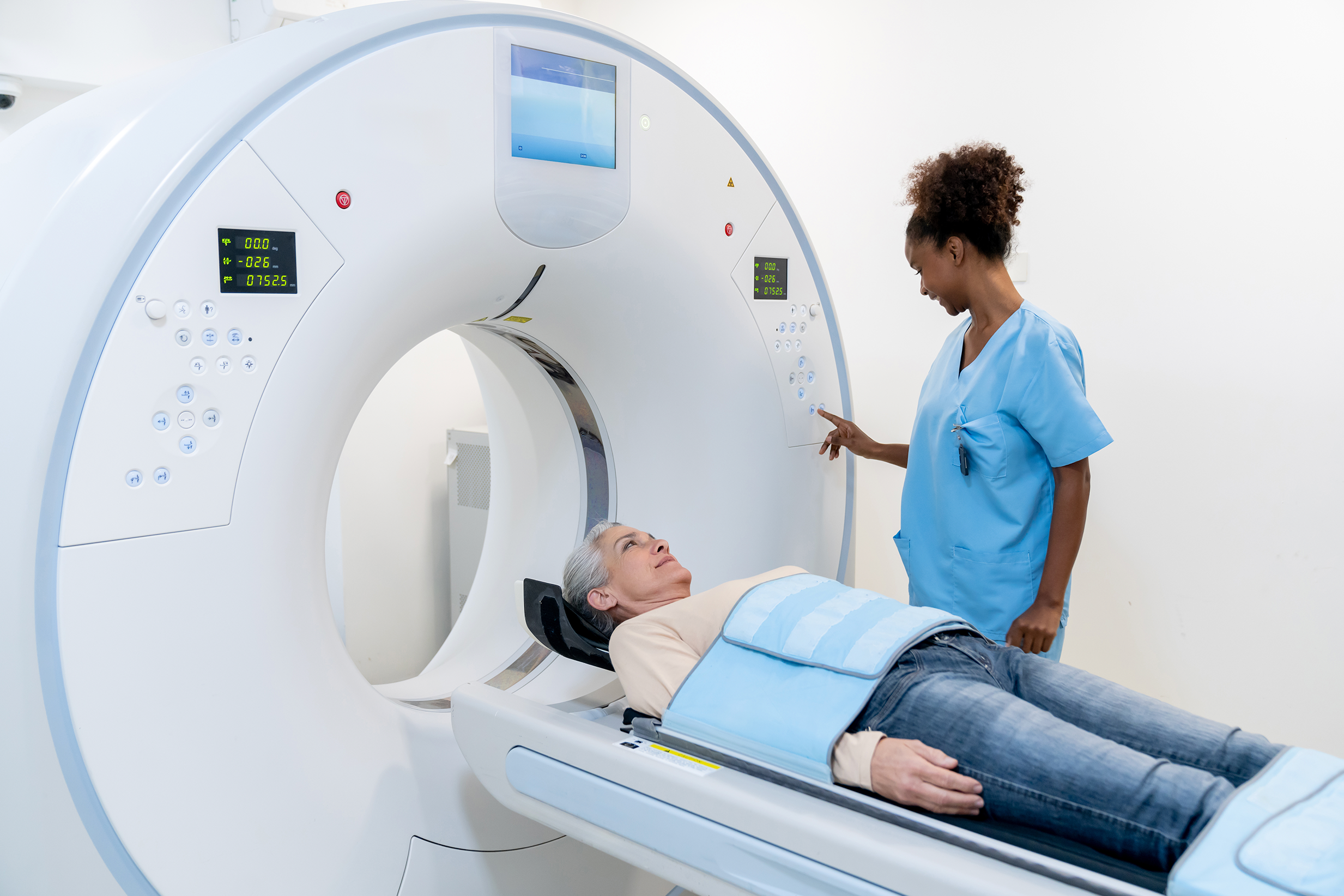
Dr. Maxine Jochelson: Yes, and it's a PET/CT, so you are getting a CT scan along with this. It's not just pure PET. So, you're seeing the bones. You're seeing all the structures, and then you're seeing the PET on top. So, yes, what we...you know, and again, there are still places that aren't using PET. There are issues in terms of insurers reimbursing for it, although it has finally made it all the way to the top in the NCCN guidelines, which is how the insurers take their cues. So, some places, they're still doing CT and bone scan, but the preponderance of the literature says that these are better tools.
So, yeah, so, if a patient comes in and they were suspecting...we're suspecting mets, then we'll do the PET or the FES, and then we'll treat the patient, and then, depending on the individual and her tumor, in two to three to four months, reimage. Lots of different people have different ways of doing it. It also depends on what drugs you're using and how the patient is...you know, if she's not responding well to the drug or if it's making her ill, they may rescan her and see what's going on, but yes.
Jamie DePolo: Okay, and the PET/CT scan together, that's done all at one time, right? You don't do one and then the other?
Dr. Maxine Jochelson: Well, you do...it's very fast. So, basically, after...we'll talk about the FDG. You've now stayed still for an hour. They'll put you on the table. They'll do the CT, but we're talking minutes. They do the CT of the whole body, and then they do the PET, and then the machine...the images are...can be read separately. So, you can read the CT with that, and then it fuses them, and so, you can then look at them...the PET on top of the CT, but the actual whole scan, the machines are getting faster and faster. So, the whole thing, from the time you lie down, you know, probably 15, 20 minutes to do the whole body. So, it's pretty quick.
Jamie DePolo: Okay. Okay, and now, kind of keeping on the metastatic lobular breast cancer, when it metastasizes, I know it tends to go to some different areas than ductal breast cancer. I want to say the gastrointestinal tract, the abdominal lining, and the tissue around the eyes or kidneys. Now, I know it doesn't go to just those areas, but those areas are a little bit different than ductal cancer. So, what kind of imaging tests do you use to kind of see those areas?
Dr. Maxine Jochelson: Yeah. Well, those are the imaging tests we've discussed.
Jamie DePolo: Oh, okay.
Dr. Maxine Jochelson: The issue is just keeping it in your mind, okay, this is invasive lobular, so I better...you know, especially the lining of the stomach, a lot of times, the metastases is just a very thin layer of cells. So, you have to look in a different way. I mean, you know, we look for everything, but you have to have, in your brain, well, in this particular patient, I really need to make sure that...it's the peritoneum, but that I take a really good look there.
You know, things like that. For every cancer that you read, you know, you kind of have an idea of the disease pattern, and then you look everywhere, but then you also go back and take a second look in those areas that are possibly more likely. And you know, the other thing, just as a note, when I talked about a genetic disorder that...where women get invasive lobular cancer, there's also a gastric cancer that goes with that, that also has the same potential genomic thing.
And actually, you can use FES on those gastric cancers, as well, because it's all coming...and it's a familial problem. You know, some families will have this. So, there is an overlap, and part of it is when the stomach is involved, diffusely involved, just like the lining. So, this is, again, you know, the reason lobular cancers are hard to see lumps, is that they grow in straight lines instead of forming a mass. They kind of insert themselves all the way through the stomach or all the way through the lining of the abdomen.
Jamie DePolo: So, it's almost like a sheet, perhaps?
Dr. Maxine Jochelson: Exactly. Exactly, and then it'll get lumpy, and there'll be more than just a thin sheet, but those are the things that we have to think about when we're looking at women with invasive lobular. Now, it will go to bones, just like other breast cancers, you know, and it will go to liver. Not as common, theoretically, but all of the regular sites can also have that cancer.
Jamie DePolo: Okay. So, just to make sure I understand, so, the testing is basically the same, but it's really the radiologist thinking, like, what is the pattern of this cancer? So, this is lobular cancer. I have to remember to look at these other areas in the scans.
Dr. Maxine Jochelson: Yes, except for the tracers that, you know...so, you wouldn't use an FES...do an FES scan on a patient with invasive ductal cancer frequently. You might if it's a low grade, but then there's HER2-positive breast cancers, and we have a completely different tracer for that, and you wouldn't use the same for both.
Jamie DePolo: Okay. Okay. Thank you for explaining that. So, I guess what I'm wondering, if somebody...and I don't know that they would, but if somebody suspected that they had lobular cancer versus ductal cancer...like, say they didn't have a lump, but maybe their breast was red, or like you said, there was some thickening, should they go to their doctor and ask for specific tests, or is the doctor going to know and say, okay, you may have lobular? We're going to do these tests?
Dr. Maxine Jochelson: Yeah. First of all, the redness in a breast is what we think of as inflammatory breast cancer, and that's not specific for lobular. That is something...it's more common to have invasive ductal, so it's more commonly seen in invasive ductal. So, the redness is not specific for that. When a patient has a breast symptom, they go to the doctor, and the doctor examines them and then orders...and you start with the routine test.
There's no reason to start with anything but your basic mammogram and ultrasound, then work up the symptoms. And then, if there is still further concern, then you can do additional testing, and many people will think about doing a breast MRI. We also have a test that we do here called contrast-enhanced mammography, and both of these are similar, in that you give women dye.
And cancer vessels are leaky, and when you give them the dye, it kind of leaks out, and it will show you something's going on in that area, even if you can't see a discreet lump, and so, if you're highly suspicious that something like that is going on with the patient and you're not finding it on the mammogram or the ultrasound, then move to...I mean, contrast mammo is relatively new, so move to MR or move to contrast to try to get a better look at what's going on.
Jamie DePolo: Okay. Okay, Dr. Jochelson, thank you so much. This has been really helpful and informative.
Dr. Maxine Jochelson: I'm glad it has. It's been interesting to think about all of these things specifically.
Jamie DePolo: Thank you.
Dr. Maxine Jochelson: You're very welcome.
Thank you for listening to The Breastcancer.org Podcast. Please subscribe on iTunes, or wherever you listen to podcasts. To share your thoughts about this or any episode, email us at podcast@breastcancer.org, or leave feedback on the podcast episode landing page on our website. And remember, you can find a lot more information about breast cancer at Breastcancer.org, and you can connect with thousands of people affected by breast cancer by joining our online community.

Affiliations: Breastlink/Radnet, Beverly Hills, Calif.
Areas of specialization: breast imaging, diagnostic radiology, nuclear radiology
Dr. Maxine Jochelson is director of mammography for Breastlink in Beverly Hills/Radnet. Besides specializing in breast imaging, she also has expertise in interpreting PET/CT scans.
Your donation goes directly to what you read, hear, and see on Breastcancer.org.