Mammograms: What You Need to Know
A mammogram is an X-ray picture of the breast tissue. Doctors use mammograms to look for abnormal changes in the breast.
Mammograms are one of the most important tools doctors have to help screen for and diagnose breast cancer. Safe and sensitive enough to pick up most breast abnormalities, the technique has been in use for more than 50 years.
Getting a mammogram is often the best way to find breast cancer early, when it’s most treatable. A mammogram can detect breast cancer before any symptoms develop. It can detect a tumor that cannot be felt. Some studies have shown that having regular mammograms can lower your risk of dying from breast cancer.
A mammogram cannot be used to diagnose breast cancer on its own, though. Other tests are always needed before a diagnosis can be made. In most cases, a biopsy is the only test that can tell for sure whether breast cancer is present.
How is a mammogram done?
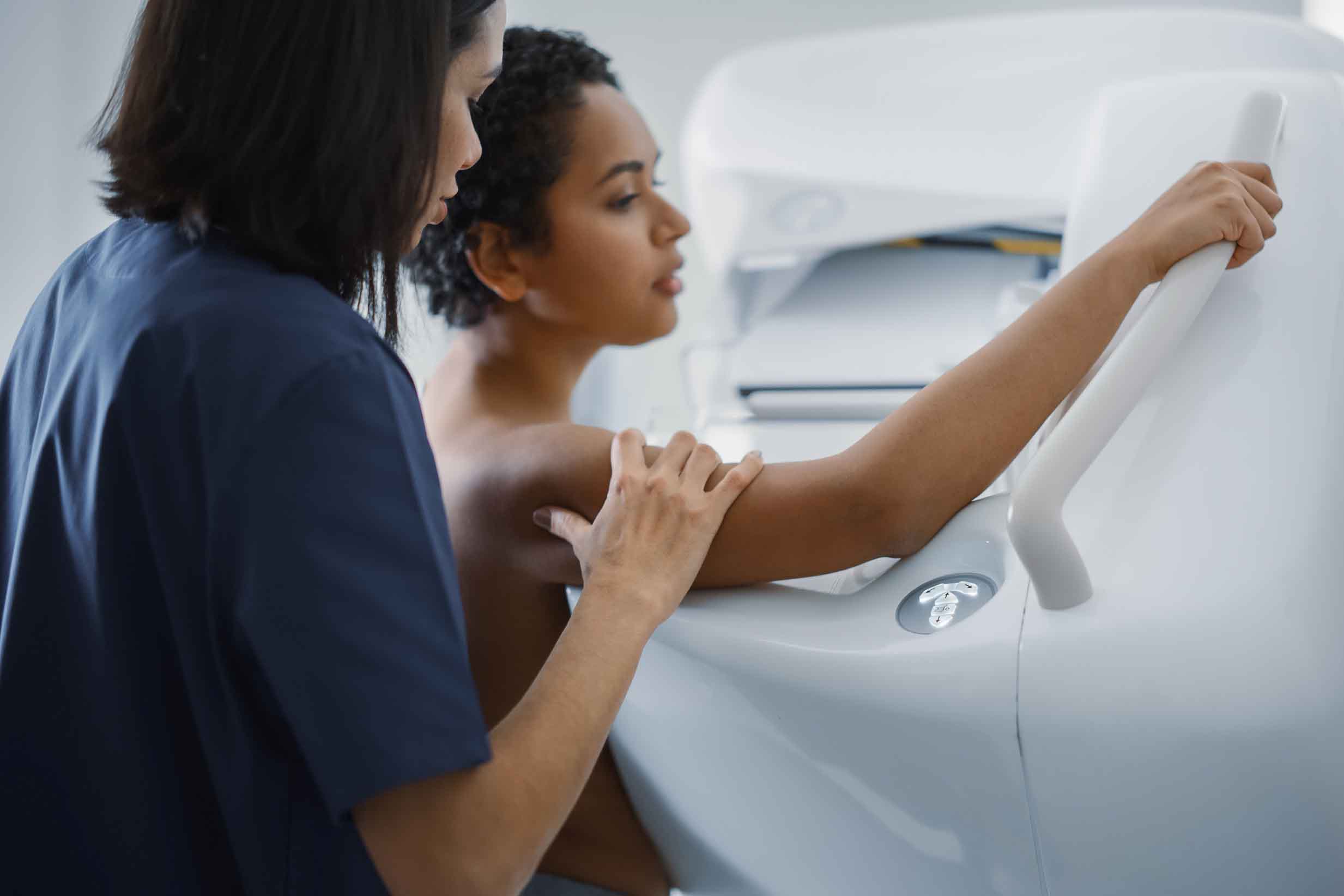
When you get a mammogram, you remove your clothing from the waist up and stand in front of a special X-ray machine. A mammogram technician positions and flattens your breast between two plastic plates. The mammogram machine takes multiple X-ray pictures of the breast tissue from different angles. Then the technician repeats the technique on the opposite breast.
The images are saved as digital files and a radiologist (a doctor who specializes in interpreting medical images) analyzes them.
Many radiologists use software called computer-aided detection (CAD) that highlights the areas on a mammogram image that may be abnormal. This can help them decide if any areas need further evaluation. Some radiologists also use artificial intelligence software to help find areas on the mammogram that may be abnormal.
Screening versus diagnostic mammograms
There are two main reasons mammograms are done, but — while the process is slightly different — the machines and the technique are the same.
Screening mammograms
Screening mammograms are used to look for signs of breast cancer in people who don’t have any symptoms of the disease. Usually two X-ray images of each breast are taken. If a potentially abnormal area in the breast is found on a screening mammogram, then your doctor will recommend that you have more tests (typically a diagnostic mammogram and an ultrasound).
Diagnostic mammograms
These are used to get more information about a specific area (or areas) of concern. They are done either because you have a possible symptom of breast cancer (such as a lump) or because a potentially abnormal area in the breast was found on a screening mammogram. A diagnostic mammogram involves taking more X-ray images, and so it usually takes longer than a screening mammogram.
Other uses for mammograms
Mammograms are also done for other reasons, such as to screen for breast cancer in people who were treated for breast cancer in the past and have some remaining breast tissue (these are sometimes called surveillance mammograms) or to see how a tumor in the breast is responding to chemotherapy that is being given before surgery (neoadjuvant chemotherapy).
Types of mammograms
Two mammogram techniques are widely used in the U.S.:
3D mammograms, also called digital breast tomosynthesis (DBT), digital tomosynthesis, or just tomosynthesis
2D digital mammograms
The experience of having 3D and 2D mammograms is similar, including the way the breast is positioned and flattened and about how long the test takes.
The main difference between the tests is the images that are produced: A 3D mammogram provides much more detail. Most doctors recommend having a 3D mammogram, if possible. Studies show that 3D mammograms find more breast cancers than 2D mammograms.
Contrast-enhanced mammograms are a special type of mammogram mostly used for follow-up imaging after something unusual is spotted on a screening mammogram.
Where to get a mammogram
You can get a high-quality mammogram at an outpatient imaging center, hospital radiology department, breast center, or mobile mammography van. A federal law requires that all mammography facilities undergo annual inspection and meet basic quality standards.
Learn more about where to get a mammogram and how to find a free or low-cost one.
How often do you need a mammogram?
Medical organizations give varying recommendations about when people with breasts should start getting routine screening mammograms and how often they should get them. But most agree that women at average risk should begin having annual mammograms at age 40, or at an earlier age if they’re at high risk for breast cancer.
If you’re unsure of your risk level, talk with your doctor about a breast cancer risk assessment. Your doctor can work with you to develop a breast cancer screening or follow-up plan that fits your risk level. It may include getting regular breast physical exams by a health care provider and getting annual mammograms. Your doctor may also recommend that you get other breast imaging tests, such as a breast MRI or ultrasound.
Learn more about mammogram screening guidelines, including who should get mammograms and when.
What mammograms show
A radiologist will look at your mammogram images to see if there are any abnormal changes in the breast tissue and if there are differences between one breast and the other. If possible, the radiologist will compare your past mammograms with your new one to check for any changes.
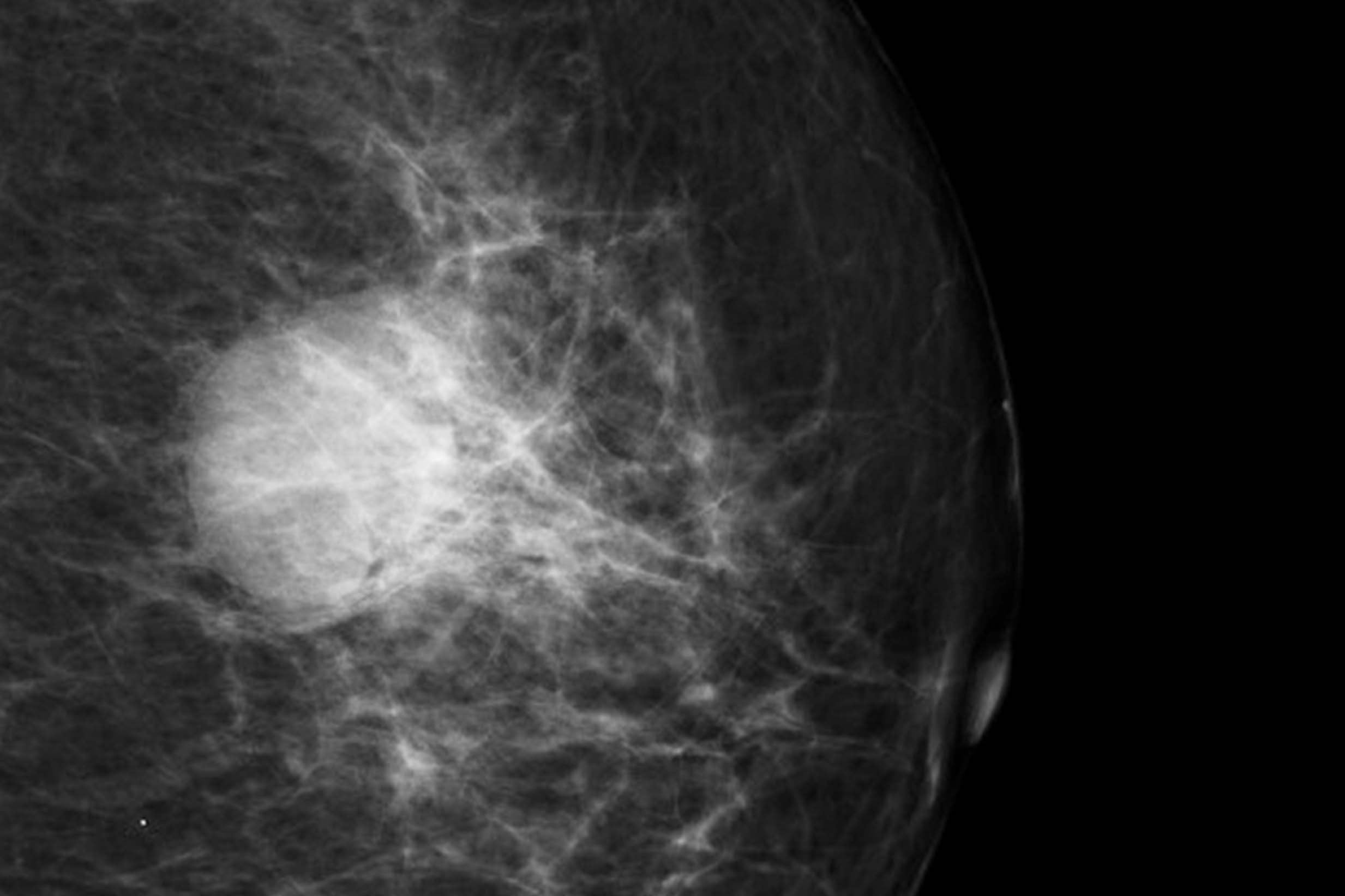
An image of a breast tumor (invasive ductal carcinoma) on a mammogram.
Courtesy of Heba Khaled Al Ja’afreh, Radiopaedia.org.
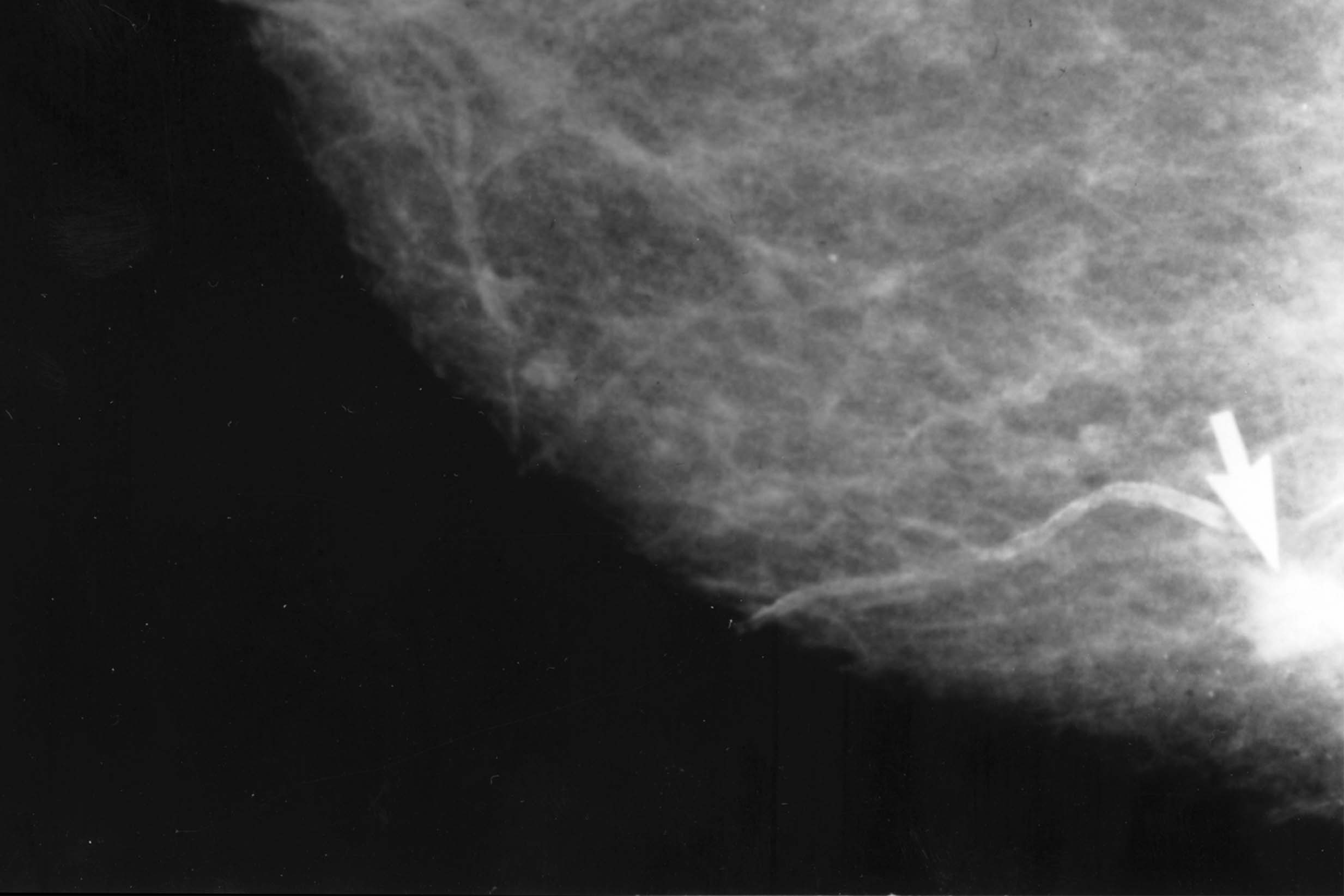
An image of an abnormal mammogram, with an arrow indicating where the tumor is.
Courtesy of Dr. Dwight Kaufman of the National Cancer Institute.
Calcifications
Calcifications are tiny flecks of the mineral calcium in the soft tissue of the breast. They’re often normal signs of aging, but can sometimes indicate the presence of an early breast cancer. Depending on how they're clustered and their shape, size, and number, your doctor may want to do further tests.
Lumps or masses
There are many types of lumps or masses, like fibroadenomas and cysts, that can be seen on a mammogram. If a mass is at least partly solid, more tests (especially an ultrasound test) may be needed to check if it could be cancerous.
Cysts and fibroadenomas are two common types of masses. Most cysts and all fibroadenomas are benign (non-cancerous).
Breast density
The radiologist who looks at your mammogram will also assess your breast density. Breast density is the amount of fibrous and glandular tissue in the breasts compared to the amount of fatty tissue in the breasts. Breast density can change over time. People with dense breast tissue have a slightly higher risk of breast cancer. Also, dense breast tissue can make it harder for a radiologist to detect breast cancer on a mammogram. If a mammogram shows that you have dense breast tissue, your doctor may recommend that you get other imaging tests (such as an ultrasound or breast MRI), too, to screen for breast cancer.
Breast asymmetry
The radiologist will check to see if the mammogram shows you have any areas of asymmetry in your breasts. An asymmetry is an area of dense breast tissue that looks different from the same area in the opposite breast.
Usually, an asymmetry is not a cause for concern. But if an asymmetry is suddenly developing or changing, it can be a sign of breast cancer or an infection and the radiologist may recommend additional testing.
How to prepare for a mammogram
When you schedule your mammogram, you may want to ask the staff at the breast imaging center about how to prepare. Here are some of the things you may need to do to get ready for your appointment.
Call your insurance company
Check with your insurance company to see what’s covered. For example, not all insurance companies fully cover a 3D mammogram, or they may cover screening mammograms at 100% but require a copay for a diagnostic mammogram. Your insurance company may also only cover a screening mammogram if a full year has passed since your last one. Checking ahead of time can help you know what costs to expect.
Find out if you need a doctor’s order
You may need a doctor’s order, especially if you’re getting a diagnostic mammogram or if you’re under 40 and getting an annual screening mammogram.
Bring your doctor’s contact info
Give the front desk staff at the breast imaging center the names and contact info for all the doctors who should receive your mammogram results. This is especially important if you want other doctors besides the one who put in the order for the mammogram to receive the results.
Ask about accommodations
If you have a disability that would make it difficult for you to stand or hold still during the mammogram, ask ahead of time about what accommodations they could provide. The technicians may be able to adjust the mammogram machine so that you can sit, for example. People with disabilities often report challenges with accommodations so calling to discuss these in advance is a good idea.
Transfer your records
If you’re going to a new breast imaging center for the first time, make sure you give them the medical records related to your previous mammograms or breast biopsies. The radiologist will want to compare your new mammogram images to your previous ones.
Talk to your doctor if you’re pregnant
If there’s any chance you might be pregnant, talk with your doctor before scheduling a mammogram or tell the staff at the breast imaging center before your mammogram. Usually, routine screening mammograms aren’t recommended for pregnant people because of the very small risk that the radiation could affect the developing fetus. However, if you found a lump or have some other potential symptom of breast cancer, your doctors may recommend that you have an ultrasound and possibly a diagnostic mammogram during pregnancy.
Learn more about mammograms during pregnancy.
Dress for comfort
Wear a top with pants, shorts, or a skirt on the day of your mammogram. Since you’ll have to undress above the waist, you may be more comfortable if you wear a two-piece outfit instead of a dress or jumpsuit.
Don’t wear deodorant or other products
Don’t wear deodorant, antiperspirant, lotion, perfume, or powder under your arms or on your chest area or breast skin on the day of your mammogram. Particles from these products can show up on your mammogram images.
Communicate with your technologist
Before your mammogram begins, let the technologist (the person operating the mammogram machine) know if...
you have breast implants. The technologist will take extra views of the breast (known as implant displacement views) if you have breast implants. This is to make sure the breast tissue is viewable on the mammogram.
you’ve recently been vaccinated. Some people who receive a COVID-19 vaccine or flu vaccine temporarily develop swollen or enlarged lymph nodes that can show up on a mammogram. The radiologist will be able to more accurately interpret your mammogram result if they know that the swelling may be from a vaccine.
you have any tattoos on your arms, chest, or back. The ink could migrate to your lymph nodes and show up as white specks on a mammogram. Learn more about how tattoo ink can show up on mammograms.
you need any accommodations.
Mammogram risks
Mammograms involve a very small amount of radiation exposure. The dose of radiation received during a screening mammogram is about the same amount a person gets from their natural surroundings (background radiation) over a period of about 7 weeks, according to the American Cancer Society. The benefits of getting regular mammograms outweigh the risks of these low doses of radiation.
The main risks of mammograms are that they may result in false negatives or false positives. A false negative means that cancer is present but isn’t found on the mammogram. Some cancers can’t be detected by mammograms. A false positive happens when an abnormality shows up on a mammogram that looks like cancer, but turns out to be benign (not cancer) after additional tests.
Do mammograms hurt?
Mammograms can be painful or at least uncomfortable. The discomfort doesn’t last long, though. Each time your breast is compressed, it will be for a few seconds at a time. And from start to finish, the entire test takes about 20 minutes. To reduce the discomfort, you can consider taking an over-the-counter pain medication about an hour before your mammogram appointment. Just be sure to check with your doctor that the pain medication doesn’t interfere with any other medications you’re taking.
What is thermography?
Thermography (also called thermal imaging) should never be used to screen for breast cancer and is not a substitute for a mammogram. This test uses an infrared camera to measure the temperature of the skin on the breast’s surface. Some health centers market it as a breast cancer screening test that can be used as an alternative to mammography, but there is no scientific evidence to support those claims, according to the FDA. Thermography can fail to detect breast cancers that other tests, such as mammograms, can detect.
Getting your mammogram results
After your mammogram, the imaging facility should send you an easy-to-understand written summary of your results. At your mammogram appointment, ask when and how you should expect to receive this summary.
In most cases, the imaging facility will ask you which of your doctors you want the results sent to. If one of your doctors put in an order for the mammogram, the facility will send them the results. The results that your doctor receives are more detailed and are based on a standardized reporting system known as BI-RADS.
Getting the results of a screening mammogram can take anywhere from 24 hours to 10 days, depending on the imaging center. The results of a diagnostic mammogram are typically given to you either during the diagnostic mammogram appointment or very soon after.
Learn more about getting your mammogram results.